Pre-Operative Management
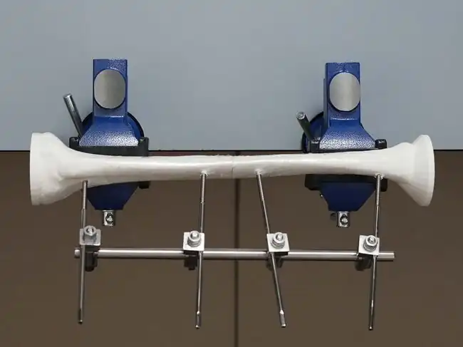
| Part of | Uniplanar External Fixation |
|---|
This module allows medical officers and surgeons who are not orthopedic specialists to become confident and competent in irrigation and debridement, powered and manual drilling, positioning and correctly inserting Schanz screws, and constructing the uniplanar external fixator frame as part of external fixation procedures for open tibial shaft fractures performed in regions without specialist coverage. To maximize patient safety, this module teaches learners to use a powered drill to insert self-drilling Schanz screws through the near cortex and then manually advance Schanz screws into the far cortex to avoid plunging.
Learning Objectives
By the end of this module, learners will be able to:
- Conduct a history and physical examination of a patient with an open tibial shaft fracture.
- Review the post-operative anteroposterior and lateral view radiographs of a patient with an open tibial shaft fracture.
- Initially manage a patient with an open tibial shaft fracture.
- Know the indications for uniplanar external fixation for a patient with an open tibial shaft fracture.
- Know the indications for referral of a patient with an open tibial shaft fracture to a tertiary center for specialist care.
History
Injury History
Date of Injury (month, day, year):___________________
Time of Injury:__________________AM;____________________PM
Date of Arrival to Hospital (month, day, year):___________________
Time of Arrival to Hospital:__________________AM;____________________PM
Mechanism of Injury
Fall
Gunshot wound
Motorcycle accident
Motor vehicle accident
Pedestrian-traffic injury
Other:___________________
Environmental Contaminants
Farmyard injury
Saltwater source
Freshwater source
Fecal contamination
Other:___________________
Allergies
Penicillin
Other:___________________
No Known Drug Allergies (NKDA)
Tetanus Vaccine Status
All patients with open fractures should be evaluated for tetanus vaccination status and receive tetanus prophylaxis in accordance with Centers for Disease Control and Prevention guidelines.[1]
| Tetanus Primary Vaccine Series Status |
> 3 doses of primary tetanus vaccine series < 3 doses Unvaccinated Unknown |
| Tetanus Booster Status |
Last dose < 5 years Last dose > 5 years Unvaccinated Unknown |
Past Medical History
Diabetes Mellitus
Immunosuppression:________________
Social History
Current smoker
Occupation:________________
Medical insurance
Physical Examination
Neurovascular Exam
Sensory Testing
| Nerve | Light Touch Sensation Testing | Sensory Exam Findings | Compare Both Sides |
|---|---|---|---|
| Lateral dorsal cutaneous branch of the sural nerve (S1-2) | Lateral aspect of the little toe |
Intact Not Intact |
Symmetric Asymmetric |
| Deep peroneal nerve (L4-5) | First dorsal webspace of the foot |
Intact Not Intact |
Symmetric Asymmetric |
| Superficial peroneal nerve (L4-S1) | Dorsum of the foot (except the first webspace) |
Intact Not Intact |
Symmetric Asymmetric |
Motor Testing
| Muscle | Motor Exam | Motor Exam Findings | Compare Both Sides |
|---|---|---|---|
| Tibialis anterior | Ankle dorsiflexion |
Able Unable |
Symmetric Asymmetric |
| Gastrocnemius and soleus | Ankle plantarflexion |
Able Unable |
Symmetric Asymmetric |
Vascular Exam
| Pulses | Vascular Exam Findings | Compare Both Sides |
|---|---|---|
| Dorsalis pedis artery: If dorsalis pedis artery pulses are not palpable, check for posterior tibial artery pulses. |
Palpable Not Palpable |
Symmetric Asymmetric |
| Posterior tibial artery |
Palpable Not Palpable |
Symmetric Asymmetric |
Acute Compartment Syndrome
Rule out acute compartment syndrome:[2]
Pain disproportionate to injury and intensified with passive stretch (i.e., flexion and extension of the toes)
Pallor
Paresthesias
Paralysis
Pulselessness
Compartment pressure greater than 30-40 mmHg in an unconscious or paralyzed patient
No signs of compartment syndrome
Photo Documentation of Injured Extremity
Take images of the soft tissue wound(s) of the injured extremity including the joint above and below for orientation.
After the wound is examined and photographed, cover it with a sterile dressing.[2]
Temporarily splint the extremity by immobilizing the joint above and below the fracture site using a backslab (plaster which does not completely encircle the limb) secured with a bandage to accommodate swelling.
Pre-Operative Open Fracture Classification
| Gustilo Type I: |
An open fracture with a wound less than 1 cm long and clean. |
| Gustilo Type II: |
An open fracture with a laceration more than 1 cm long without extensive soft tissue damage, flaps, or avulsions. |
| Gustilo Type IIIA: |
Adequate soft-tissue coverage of a fractured bone despite extensive soft-tissue laceration or flaps, or high-energy trauma irrespective of the size of the wound. |
| Gustilo Type IIIB: |
Extensive soft-tissue injury loss with periosteal stripping and bone exposure. This is usually associated with massive contamination. |
| Gustilo Type IIIC: |
Open fracture associated with arterial injury requiring repair. |
Preoperative Radiographic Findings
Review anteroposterior (AP) and lateral views and note any clinically significant radiographic findings.
| X-Ray Feature | Check the most appropriate response |
|---|---|
| Injured lower extremity side |
Left Right |
| Tibial fracture site |
Diaphysis (shaft) Metaphysis Epiphysis |
| Tibial fracture location |
Proximal 1/3 Middle 1/3 Distal 1/3 |
| Joint involvement |
Extra-articular Intra-articular |
| Tibial fracture pattern |
Transverse Oblique Spiral Comminuted (> 2 fragments) Segmental Other:___________________ |
| Tibial fracture bone apposition |
> 50% bone apposition[5] < 50% bone apposition |
| Tibial fracture angulation should be assessed in the coronal and sagittal plane. The anteroposterior view shows the coronal plane and the lateral view shows the sagittal plane. |
< 10 degrees of angulation in the coronal or saggital plane[6][7][8] >10 degrees of angulation in the coronal or saggital plane |
| Fibular fracture site |
Diaphysis (shaft) Metaphysis Epiphysis None |
Indications for Uniplanar External Fixation
After completing the entire module, learners should be able to perform uniplanar external fixation of open tibial shaft fractures with all of the following features:
Able to directly visualize fracture through the open wound or intraoperative extension of the wound; and
Gustilo Type II or Gustilo Type IIIA open tibial fracture; and
Non-comminuted, non-segmental tibial shaft (extra-articular) fracture; and
With or without a fibular shaft (extra-articular) fracture
Indications for Referral to a Tertiary Center for Specialist Care
The learner should refer a patient with any of the following features to a tertiary center for specialist care:
Unable to directly visualize fracture through the open wound or intraoperative extension of the wound
Non-palpable pedal pulse
Symptoms consistent with acute compartment syndrome
Gustilo Type IIIB or Gustilo Type IIIC open tibial fracture
Comminuted or segmental tibial fracture
Bilateral tibia fractures
Metaphyseal tibial fracture with intra-articular extension
Concomitant distal fibular fracture near or involving the ankle joint
Concomitant ipsilateral or contralateral femoral fracture
Severe traumatic brain injury (Glasgow Coma Scale <12)
Severe spinal cord injury (lower extremity paresis/paralysis)
Severe burns (involving >10% of the total body surface area or >5% of the total body surface area with full-thickness or circumferential injury)
Fracture Management Plan
Advanced Trauma Life Support
Any patient with a fracture should be initially managed as a trauma patient using Advanced Trauma Life Support protocols (life-threatening conditions treated first).
Diet
Nil per os (NPO) 6 hours prior to surgery
Laboratory Testing
Type and screen (group and crossmatching)
CBC (hematocrit, platelets)
Glucose
Potassium
Creatinine
Additional pre-operative investigations may be required depending on patient factors.
Antibiotic Therapy
All patients will be managed with intravenous antibiotics immediately at the time of presentation to the emergency department.[9][10][11] Antibiotics may be changed, added or extended depending on subsequent clinical findings. Doses will be adjusted based on patient weight when indicated.
Recommended Antibiotic Therapies for Open Fractures
| Injury Characteristics | Systemic Antibiotic Regimen | Penicillin Allergy |
|---|---|---|
| Gustilo Type I and II | Cefazolin 2 g IV immediately and q8 hours for a total of 3 doses[9][10][11] | Clindamycin 900 mg IV immediately and q8 hours for a total of 3 doses |
| Gustilo Type III |
Ceftriaxone 2 g IV immediately for a total of 1 dose, and Vancomycin 1 g IV immediately and q12 hours for a total of 2 doses |
Aztreonam 2 g IV immediately and q8 hours for a total of 3 doses, and Vancomycin 1 g IV immediately and q12 hours for a total of 2 doses |
| Farm or fecal
contamination |
Add Penicillin G IV (e.g., 5 million-10 million units/24 hours)[9][10] | Add Metronidazole IV |
| Freshwater or
saltwater contamination |
Add Levofloxacin IV or Ciprofloxacin IV[11] | Add Levofloxacin IV or Ciprofloxacin IV[11] |
Tetanus Prophylaxis
All patients with open fractures should receive tetanus prophylaxis in accordance with Centers for Disease Control and Prevention guidelines.[1]
Tetanus toxoid booster; or
Tetanus toxoid primary series; or
250 IU tetanus immune globulin IM; or
Not required
Acknowledgements
This work is funded by a grant from the Intuitive Foundation. Any research, findings, conclusions, or recommendations expressed in this work are those of the author(s), and not of the Intuitive Foundation.
References
- ↑ 1.0 1.1 https://www.cdc.gov/tetanus/clinicians.html
- ↑ 2.0 2.1 Berg, E.E. and Murnaghan, J.J. Orthopedic Surgery: Diseases of the Musculoskeletal System. Essentials of surgical specialties, 2nd Edition. Edited by Peter F Lawrence. 514 pages, illustrated. Philadelphia: Lippincott Williams & Wilkins, 2000.
- ↑ Gustilo RB, Anderson JT. Prevention of infection in the treatment of one thousand and twenty-five open fractures of long bones: retrospective and prospective analyses. J Bone Joint Surg Am. 1976 Jun;58(4):453-8. PMID:773941.
- ↑ Gustilo RB, Mendoza RM, Williams DN. Problems in the management of type III (severe) open fractures: a new classification of type III open fractures. J Trauma.1984 Aug;24(8):742-6. doi: 10.1097/00005373-198408000-00009. PMID:6471139.
- ↑ https://www.orthobullets.com/trauma/1045/tibial-shaft-fractures
- ↑ Nicoll EA. Fractures of the tibial shaft. A survey of 705 cases. J Bone Joint Surg Br. 1964 Aug;46:373-87.
- ↑ Haonga BT, Liu M, Albright P, Challa ST, Ali SH, Lazar AA, Eliezer EN, Shearer DW, Morshed S. Intramedullary Nailing Versus External Fixation in the Treatment of Open Tibial Fractures in Tanzania: Results of a Randomized Clinical Trial. J Bone Joint Surg Am. 2020 May 20;102(10):896-905. doi: 10.2106/JBJS.19.00563. PMID: 32028315; PMCID: PMC7508278.
- ↑ Merchant TC, Dietz FR. Long-term follow-up after fractures of the tibial and fibular shafts. J Bone Joint Surg Am. 1989 Apr;71(4):599-606. PMID: 2703519.
- ↑ 9.0 9.1 9.2 Garner MR, Sethuraman SA, Schade MA, Boateng H. Antibiotic Prophylaxis in Open Fractures: Evidence, Evolving Issues, and Recommendations. J Am Acad Orthop Surg. 2020 Apr 15;28(8):309-315. doi: 10.5435/JAAOS-D-18-00193. PMID: 31851021.
- ↑ 10.0 10.1 10.2 https://surgeryreference.aofoundation.org/orthopedic-trauma/adult-trauma/tibial-shaft/further-reading/principles-of-management-of-open-fractures?searchurl=%2fSearchResults#principles-of-surgical-care-for-open-fractures
- ↑ 11.0 11.1 11.2 11.3 Zhu H, Li X, Zheng X. A Descriptive Study of Open Fractures Contaminated by Seawater: Infection, Pathogens, and Antibiotic Resistance. Biomed Res Int. 2017;2017:2796054. doi: 10.1155/2017/2796054. Epub 2017 Feb 20. PMID: 28303249; PMCID: PMC5337837.
| Authors | Medical Makers, Julielynn Wong |
|---|---|
| License | CC-BY-SA-4.0 |
| Cite as | Medical Makers, Julielynn Wong (2022–2025). "Uniplanar External Fixation/Knowledge Review/Pre-Operative Management". Appropedia. Retrieved November 28, 2025. |