Post-Operative Management
| Part of | Uniplanar External Fixation |
|---|
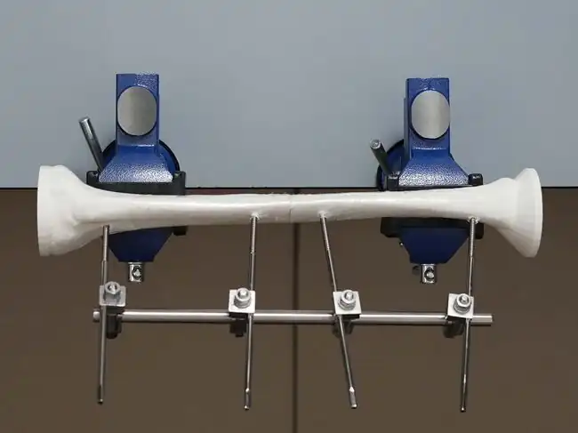
This module allows medical officers and surgeons who are not orthopedic specialists to become confident and competent in irrigation and debridement, powered and manual drilling, positioning and correctly inserting Schanz screws, and constructing the uniplanar external fixator frame as part of external fixation procedures for open tibial shaft fractures performed in regions without specialist coverage. To maximize patient safety, this module teaches learners to use a powered drill to insert self-drilling Schanz screws through the near cortex and then manually advance Schanz screws into the far cortex to avoid plunging.
Learning Objectives
By the end of this module, learners will be able to:
- Conduct a history and physical examination of a patient following uniplanar external fixation of an open tibial shaft fracture.
- Review the post-operative anteroposterior and lateral view radiographs of a patient following uniplanar external fixation of an open tibial shaft fracture.
- Provide post-operative care for a patient following uniplanar external fixation of an open tibial shaft fracture.
- Know the indications for adjusting the uniplanar external fixator for a patient with an open, simple tibial shaft fracture.
- Know the indications for referral of a patient with an open tibial shaft fracture to a tertiary center for specialist care.
Postoperative Clinical Assessment
Physical Examination
| Clinical Issue | Clinical Signs | Check the most appropriate response | Management |
|---|---|---|---|
| Highly contaminated wound | Post-operative wound has non-viable tissue | Yes
No |
If the post-operative wound is still contaminated, additional debridement procedures can be performed 48 to 72 hours apart until the wound is clean. |
| Fracture-related infection |
Fistula, sinus or wound breakdown (with communication to the bone or the implant); or Purulent drainage from the wound[1] |
Yes No |
If there are clinical signs of a fracture-related infection, the patient should be referred to a tertiary center for specialist care. |
| Skin tenting |
Skin tension around any of 4 pin sites ("when skin tracks up pin")[2] |
Yes No |
If skin tenting is present, the stab incision should be widened to release any soft tissue tension around the pin site to reduce the risk of inflammation and pin infection.[3] |
| Patellar ligament tethering |
"Far" Schanz screw in the proximal fragment is placed directly on or proximal to the tibial tuberosity, and Limited range of motion for knee flexion due to localized pain |
Yes No |
If there is patellar ligament tethering, the pin must be changed. The patient should return to the operating room for pin re-positioning medial or distal to the tibial tuberosity. |
Neurovascular Exam
Sensory Testing
| Nerve | Light Touch Sensation Testing | Sensory Exam Findings | Compare Both Sides |
|---|---|---|---|
| Lateral dorsal cutaneous branch of the sural nerve (S1-2) | Lateral aspect of the little toe |
Intact Not Intact |
Symmetric Asymmetric |
| Deep peroneal nerve (L4-5) | First dorsal webspace of the foot |
Intact Not Intact |
Symmetric Asymmetric |
| Superficial peroneal nerve (L4-S1) | Dorsum of the foot (except the first webspace) |
Intact Not Intact |
Symmetric Asymmetric |
Motor Testing
| Muscle | Motor Exam | Motor Exam Findings | Compare Both Sides |
|---|---|---|---|
| Tibialis anterior | Ankle dorsiflexion |
Able Unable |
Symmetric Asymmetric |
| Gastrocnemius and soleus | Ankle plantarflexion |
Able Unable |
Symmetric Asymmetric |
Vascular Exam
| Pulses | Vascular Exam Findings | Compare Both Sides |
|---|---|---|
| Dorsalis pedis artery: If dorsalis pedis artery pulses are not palpable, check for posterior tibial artery pulses. |
Palpable Not Palpable |
Symmetric Asymmetric |
| Posterior tibial artery |
Palpable Not Palpable |
Symmetric Asymmetric |
Acute Compartment Syndrome
Rule out acute compartment syndrome:[4]
Pain disproportionate to injury and intensified with passive stretch (i.e., flexion and extension of the toes)
Pallor
Paresthesias
Paralysis
Pulselessness
Compartment pressure greater than 30-40 mmHg in an unconscious or paralyzed patient
No signs of compartment syndrome
Hardware Inspection
| Hardware Issue | Clinical Signs | Check the most appropriate response | Management |
|---|---|---|---|
| Pin site infection | Cellulitis around the pin site (i.e., local erythema, swelling, tenderness)
Discharge from the pin site |
Yes No |
Pin site infections are not considered surgical site infections.[5] If a pin site infection is present, the pin site should be cleaned daily and a short course of antibiotics may be given. Pin site infections do not require surgical removal of pin unless the infection persists and leads to pin loosening. |
| Pin loosening | Pain around the pin site
Seropurulent discharge from the pin site[6] Pin can be freely turned by hand when the pin-to-rod clamp is loosened |
Yes No |
Normally, a pin cannot be turned by hand when the pin-to-rod clamp is loosened. If a pin site infection persists leading to pin loosening, the pin should be removed in the operating room, > two deep tissue specimens (i.e., pin tract curettes) should be taken to obtain wound cultures, and a new pin should be inserted 2.0 cm from the original pin site.[1] |
| Pin failure | Broken pin(s) |
Yes No |
A broken pin should be removed in the operating room and a new pin inserted 2.0 cm from the original pin site. |
| Frame loosening | Clamp(s) not securely tightened | Yes
No |
Use the 11 mm spanner with T handle to tighten the clamps at the bedside. |
| Frame component failure | Broken clamp(s) or rod | Yes
No |
Replace the failed component at the bedside and notify the hardware manufacturer. |
Other Clinical Findings
Please describe any other relevant clinical findings:______________________________________
Photo Documentation of Injured Extremity
Take images of the soft tissue wound(s) of the injured extremity including the joint above and below for orientation.
After the wound is examined and photographed, re-dress it with a sterile dressing.
Post-Operative Radiographic Findings
Review anteroposterior (AP) and lateral views and note any clinically significant radiographic findings.[7] The AP view shows the coronal plane and the lateral view shows the sagittal plane.
| X-Ray Feature | Check the most appropriate response | Management |
|---|---|---|
| Adequate tibial fracture reduction
> 50% bone apposition;[8] or < 10 degrees of angulation in the coronal or sagittal plane[5][9][10] |
Yes No |
If the fracture reduction is inadequate, an additional procedure can be performed 48 to 72 hours after the initial procedure to achieve fracture alignment within acceptable parameters |
| Pin within fracture line |
Yes No |
Surgical removal and replacement of pin placed at least 2.0 cm from the fracture line |
| Pin site fracture |
Yes No |
If a fracture around a pin occurs, refer patient to a tertiary center for specialist care. |
| Pin entry in joint |
Pin in knee joint Pin in ankle joint Pin not in joint |
Surgical removal and re-insertion of a new pin outside of the joint |
Post-Operative Management Plan
Wound Care
Soft tissue wounds are cleaned with sterile normal saline and dressed with povidone iodine gauze dressing.
The frequency of dressing changings will vary according to the amount of wound contamination:
If the wound is highly contaminated, dressings are changed twice daily.
If the wound is clean, dressings can be changed once daily or every other day.
Antibiotic Therapy
Antibiotics may be changed, added or extended depending on clinical findings.[11][12][13] Doses will be adjusted based on patient weight when indicated.
Recommended Antibiotic Therapies for Open Fractures
| Injury Characteristics | Systemic Antibiotic Regimen | Penicillin Allergy | |
|---|---|---|---|
| Gustilo Type I and II |
Cefazolin 2 g IV immediately and q8 hours for a total of 3 doses[11][12][13] |
Clindamycin 900 mg IV immediately and q8 hours for a total of 3 doses | |
| Gustilo Type III |
Ceftriaxone 2 g IV immediately for a total of 1 dose, and Vancomycin 1 g IV immediately and q12 hours for a total of 2 doses |
Aztreonam 2 g IV immediately and q8 hours for a total of 3 doses, and Vancomycin 1 g IV immediately and q12 hours for a total of 2 doses | |
| Farm or fecal
contamination |
Add Penicillin G IV (e.g., 5 million-10 million units/24 hours)[11][13] |
Add Metronidazole IV | |
| Freshwater or
saltwater contamination |
Add Levofloxacin IV or Ciprofloxacin IV[12] |
Add Levofloxacin IV or Ciprofloxacin IV[12] |
Venous Thromboembolism Prophylaxis
Contraindications for venous thromboembolism (VTE) chemoprophylaxis are:[14]
active bleeding in the last 48–72 hours
hypertensive crisis
coagulopathy
platelet count < 25,000
used recombinant tissue plasminogen activator against stroke within 24 hours
recent head trauma with central nervous system hemorrhage
multiple trauma with high bleeding risk, such as solid organ injury (suspected) peri-spinal hematoma, or
at high risk for bleeding according to clinical judgment
if no contraindications for VTE chemoprophylaxis, give enoxaparin (low molecular weight heparin) 40 mg every 24 h
Early Mobilization
On post-operative day #2, the patient can be non-weightbearing and mobilized on axillary crutches.
Additional Surgical Procedures
If required, additional procedures can be performed 48 to 72 hours later for further debridement (a "second look") if the wound is still contaminated, suture-based skin closure of a clean, small (< 4 cm) soft tissue wound, and/or to adjust or replace the uniplanar external fixator hardware.
Post-operative AP and lateral radiographs will be obtained and reviewed.
Antibiotics may be changed, added or extended depending on clinical findings.
Discharge Instructions
When the wound appears clean enough for dressing changes twice a week, patients will remain non-weight-bearing, and can be discharged typically within 48 hours on crutches with wound and pin care instructions.
The patient or caregiver should apply the following wound and pin care instructions until the uniplanar external fixator is removed:
- Clean soft tissue wounds should be dressed with a non-adherent gauze dressing (like Sofra-Tulle) twice a week.
- The pin insertion sites normally do not have to be dressed. However, if a clean environment and hygiene cannot be maintained after discharge, then gauze soaked in povidone iodine can be used to dress the pin insertion sites.
- The pin insertion sites should be kept clean. If crust or exudate is present, then the pin insertion site can be cleaned with normal saline and disinfected with alcohol.
- Pin insertion sites need not be protected for showering or bathing with clean water.[15]
Follow-up Care
After discharge, patients can be followed up in the clinic at 2 weeks, 4 weeks, 6 weeks, 8 weeks, 12 weeks, 6 months, and 1 year or as per local practices.
Acknowledgements
This work is funded by a grant from the Intuitive Foundation. Any research, findings, conclusions, or recommendations expressed in this work are those of the author(s), and not of the Intuitive Foundation.
References
- ↑ 1.0 1.1 Metsemakers WJ, Morgenstern M, McNally MA, Moriarty TF, McFadyen I, Scarborough M, Athanasou NA, Ochsner PE, Kuehl R, Raschke M, Borens O, Xie Z, Velkes S, Hungerer S, Kates SL, Zalavras C, Giannoudis PV, Richards, RG, Verhofstad MHJ. Fracture-related infection: A consensus on definition from an international expert group. Injury. 2018 Mar;49(3):505-510. doi:10.1016/j.injury.2017.08.040. Epub 2017 Aug 24. PMID: 28867644.
- ↑ https://www.wacountry.health.wa.gov.au/~/media/WACHS/Documents/About-us/Policies/Pin-Site-Care-Addendum.PDF?thn=0
- ↑ https://surgeryreference.aofoundation.org/orthopedic-trauma/adult-trauma/tibial-shaft/simple-fracture-transverse/modular-external-fixator#aftercare-following-external-fixation
- ↑ Berg, E.E. and Murnaghan, J.J. Orthopedic Surgery: Diseases of the Musculoskeletal System. Essentials of surgical specialties, 2nd Edition. Edited by Peter F Lawrence. 514 pages, illustrated. Philadelphia: Lippincott Williams & Wilkins, 2000.
- ↑ 5.0 5.1 Haonga BT, Liu M, Albright P, Challa ST, Ali SH, Lazar AA, Eliezer EN, Shearer DW, Morshed S. Intramedullary Nailing Versus External Fixation in the Treatment of Open Tibial Fractures in Tanzania: Results of a Randomized Clinical Trial. J Bone Joint Surg Am. 2020 May 20;102(10):896-905. doi:10.2106/JBJS.19.00563. PMID: 32028315; PMCID: PMC7508278.
- ↑ Encinas-Ullán CA, Martínez-Diez JM, Rodríguez-Merchán EC. The use of external fixation in the emergency department: applications, common errors, complications and their treatment. EFORT Open Rev. 2020 Apr 2;5(4):204-214. doi: 10.1302/2058-5241.5.190029. PMID: 32377388; PMCID: PMC7202044.
- ↑ Ibrahim J, Liu M, Yusi K, Haonga B, Eliezer E, Shearer DW, Morshed S. Conducting a Randomized Controlled Trial in Tanzania: Institute for Global Orthopaedics and Traumatology and the Muhimbili Orthopaedic Institute. J Orthop Trauma. 2018 Oct;32 Suppl 7:S47-S51. doi:10.1097/BOT.0000000000001294. PMID: 30247401.
- ↑ https://www.orthobullets.com/trauma/1045/tibial-shaft-fractures
- ↑ Nicoll EA. Fractures of the tibial shaft. A survey of 705 cases. J Bone Joint Surg Br. 1964 Aug;46:373-87.
- ↑ Merchant TC, Dietz FR. Long-term follow-up after fractures of the tibial and fibular shafts. J Bone Joint Surg Am. 1989 Apr;71(4):599-606. PMID: 2703519.
- ↑ 11.0 11.1 11.2 Garner MR, Sethuraman SA, Schade MA, Boateng H. Antibiotic Prophylaxis in Open Fractures: Evidence, Evolving Issues, and Recommendations. J Am Acad Orthop Surg. 2020 Apr 15;28(8):309-315. doi: 10.5435/JAAOS-D-18-00193. PMID: 31851021.
- ↑ 12.0 12.1 12.2 12.3 Zhu H, Li X, Zheng X. A Descriptive Study of Open Fractures Contaminated by Seawater: Infection, Pathogens, and Antibiotic Resistance. Biomed Res Int. 2017;2017:2796054. doi: 10.1155/2017/2796054. Epub 2017 Feb 20. PMID: 28303249; PMCID: PMC5337837.
- ↑ 13.0 13.1 13.2 https://surgeryreference.aofoundation.org/orthopedic-trauma/adult-trauma/tibial-shaft/further-reading/principles-of-management-of-open-fractures?searchurl=%2fSearchResults#principles-of-surgical-care-for-open-fractures
- ↑ Gunning AC, Maier RV, de Rooij D, Leenen LPH, Hietbrink F. Venous thromboembolism (VTE) prophylaxis in severely injured patients: an international comparative assessment. Eur J Trauma Emerg Surg. 2021 Feb;47(1):137-143. doi: 10.1007/s00068-019-01208-z. Epub 2019 Aug 30. PMID: 31471670; PMCID: PMC7851035.
- ↑ https://surgeryreference.aofoundation.org/orthopedic-trauma/adult-trauma/tibial-shaft/simple-fracture-transverse/modular-external-fixator#aftercare-following-external-fixation
| Authors | Medical Makers, Julielynn Wong |
|---|---|
| License | CC-BY-SA-4.0 |
| Cite as | Medical Makers, Julielynn Wong (2022–2025). "Uniplanar External Fixation/Knowledge Review/Post-Operative Management". Appropedia. Retrieved November 28, 2025. |