This module allows traditional bone setters, pre-hospital providers, clinical officers, nurses, nurse practitioners, and medical officers to become confident and competent in performing point-of-care ultrasound diagnostic imaging to rule out the presence of a pediatric distal forearm fracture and distinguish between buckle (torus) fractures and cortical break fractures to make appropriate referrals as part of the management of closed pediatric (< 16 years of age) distal forearm fractures in regions without access to X-ray imaging and orthopedic specialist coverage.[1][2][3][4][5][6][7][8][9]
Anatomy Review
Learning Objectives
Identify the dorsal, volar (palmar), radial and ulnar surfaces of the forearm
Identify the proximal and distal regions of the forearm
Identify the radius and ulna bones of the forearm
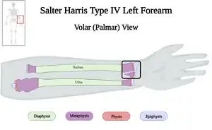
Identify the diaphysis (shaft), metaphysis, physis (growth plate) and epiphysis of the long bones of the juvenile forearm
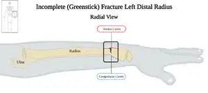
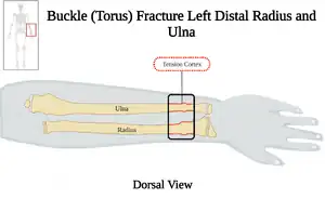
Identify the tension and compression cortex of the radius and ulna for buckle (torus) and incomplete (greenstick) fractures
Describe the differences between the pediatric distal forearm fracture subtypes, including; buckle (torus), incomplete (greenstick), complete and physeal (Salter-Harris) fractures
Recognize no fracture, buckle (torus) fracture, cortical breach fracture of the pediatric distal forearm, and other fractures of the forearm
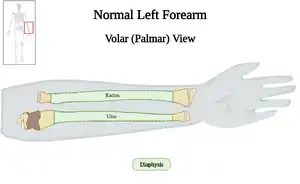
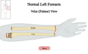
Surface Anatomy of the Forearm
Surface Anatomy of the Forearm
#
Surface
Reference Image
1
Dorsal
2
Volar (Palmar)
3
Radial
4
Ulnar
5
Proximal and Distal
Long Bone Anatomy
Long Bone Anatomy
#
Anatomic Structure
Description
Reference Image
1
Diaphysis
Body (shaft) of bone
2
Metaphysis
Flared part of bone shaft
3
Physis
Growth plate made of cartilage; also known as epiphyseal plate
4
Epiphysis
End of bone
Classification of Pediatric Distal Forearm Fractures
This work is funded by a grant from the Intuitive Foundation. Any research, findings, conclusions, or recommendations expressed in this work are those of the author(s), and not of the Intuitive Foundation.
References
↑Onyemaechi NO, Itanyi IU, Ossai PO, Ezeanolue EE. Can traditional bonesetters become trained technicians? Feasibility study among a cohort of Nigerian traditional bonesetters. Hum Resour Health. 2020 Mar 20;18(1):24. doi: 10.1186/s12960-020-00468-w. PMID: 32197617; PMCID: PMC7085192.
↑Heiner JD, McArthur TJ. The ultrasound identification of simulated long bone fractures by prehospital providers. Wilderness Environ Med. 2010 Jun;21(2):137-40. doi: 10.1016/j.wem.2009.12.028. Epub 2009 Dec 22. PMID: 20591377.
↑Heiner JD, Baker BL, McArthur TJ. The ultrasound detection of simulated long bone fractures by U.S. Army Special Forces Medics. J Spec Oper Med. 2010 Spring;10(2):7-10. PMID: 20936597.
↑Heiner JD, Proffitt AM, McArthur TJ. The ability of emergency nurses to detect simulated long bone fractures with portable ultrasound. Int Emerg Nurs. 2011 Jul;19(3):120-4. doi: 10.1016/j.ienj.2010.08.004. Epub 2010 Sep 25. PMID: 21665155.
↑Snelling PJ, Jones P, Keijzers G, Bade D, Herd DW, Ware RS. Nurse practitioner administered point-of-care ultrasound compared with X-ray for children with clinically non-angulated distal forearm fractures in the ED: a diagnostic study. Emerg Med J. 2021 Feb;38(2):139-145. doi: 10.1136/emermed-2020-209689. Epub 2020 Sep 8. PMID: 32900856.
↑Snelling PJ, Jones P, Moore M, Gimpel P, Rogers R, Liew K, Ware RS, Keijzers G. Describing the learning curve of novices for the diagnosis of paediatric distal forearm fractures using point-of-care ultrasound. Australas J Ultrasound Med. 2022 Mar 7;25(2):66-73. doi: 10.1002/ajum.12291. PMID: 35722050; PMCID: PMC9201201.
↑Heiner JD, McArthur TJ. A simulation model for the ultrasound diagnosis of long-bone fractures. Simul Healthc. 2009 Winter;4(4):228-31. doi: 10.1097/SIH.0b013e3181b1a8d0. PMID: 19915442.
↑Snelling PJ, Keijzers G, Byrnes J, Bade D, George S, Moore M, Jones P, Davison M, Roan R, Ware RS. Bedside Ultrasound Conducted in Kids with distal upper Limb fractures in the Emergency Department (BUCKLED): a protocol for an open-label non-inferiority diagnostic randomised controlled trial. Trials. 2021 Apr 14;22(1):282. doi: 10.1186/s13063-021-05239-z. PMID: 33853650; PMCID: PMC8048294.
↑Snelling PJ. A low-cost ultrasound model for simulation of paediatric distal forearm fractures. Australas J Ultrasound Med. 2018 Feb 25;21(2):70-74. doi: 10.1002/ajum.12083. PMID: 34760505; PMCID: PMC8409885.
↑Light TR, Ogden DA, Ogden JA: The anatomy of metaphyseal torus fractures. Clin Orthop Relat Res 1984, 103-111.
↑ 12.012.112.212.312.412.512.612.7Randsborg PH, Sivertsen EA. Classification of distal radius fractures in children: good inter- and intraobserver reliability, which improves with clinical experience. BMC Musculoskelet Disord. 2012 Jan 23;13:6. doi: 10.1186/1471-2474-13-6. PMID: 22269925; PMCID: PMC3331853.
↑Dietzel M, Scherer S, Esser M, Kirschner HJ, Fuchs J, Lieber J. Fractures of the proximal radius in children: management and results of 100 consecutive cases. Arch Orthop Trauma Surg. 2022 Aug;142(8):1903-1910. doi: 10.1007/s00402-021-03917-w. Epub 2021 May 11. PMID: 33974141; PMCID: PMC9296417.
↑Levine RH, Foris LA, Nezwek TA, et al. Salter Harris Fractures. [Updated 2022 Apr 21]. In: StatPearls [Internet]. Treasure Island (FL): StatPearls Publishing; 2022 Jan-. Available from: https://www.ncbi.nlm.nih.gov/books/NBK430688/
_Fracture_of_Pediatric_Female_Left_Distal_Radius_v2.0.jpg)